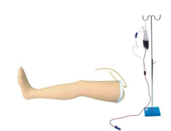
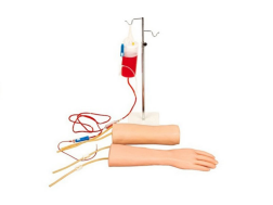
Reference product link:https://www.yddol.com/products/74.html
Introduction
The childbirth mechanism model is a valuable educational tool both in teaching and in patient care during delivery. The accompanying manual provides general guidance on obstetric techniques, including:
Palpation of fetal head, shoulders, back, knees, and elbows
Normal vaginal delivery, cesarean section, breech presentation, single-footling breech, and Ritgen's maneuver
Multiple pregnancies, including: double-head, head-breech, breech-head, double breech, and cord prolapse
Placenta previa: complete, partial, and marginal fetal suction (can be done in any position)
This model allows the placement of the fetus in any position to simulate childbirth scenarios, with more anatomical landmarks on a life-size pelvis. The peritoneal cavity is open, and the flexible abdominal "cover" can be removed. The birth canal size is normal, and the vulva and perineum are made of soft materials, which can be replaced if needed. The model provides two fetal models for practicing palpation and various fetal positions during birth. Each fetus is about 19 inches long, with specific markers such as: the fontanel (on the soft part of the head, indicating unfused skull bones), nose, mouth, ears, and tactile sensations to the spine. The fetus has a blue cord resembling the umbilical vein and two umbilical arteries, with a red placenta and umbilical cord separated by Velcro within the maternal abdominal wall. The placenta can be fixed in various positions to simulate different placental placements. A note: when simulating delivery, large amounts of talcum powder are applied to the fetal shoulders to ensure smooth delivery and easier cleaning of the model afterward.
Cesarean Section
A cesarean section (C-section) involves an incision through the abdominal wall and uterus to deliver the fetus. C-sections are often performed due to breech position, prolonged labor, labor weakness, fetal distress, cord prolapse, placenta previa, placental abruption, or other abnormalities.
The C-section incision can be horizontal or vertical. A low transverse incision is a narrow horizontal cut below the belly button, while a vertical incision is made between the navel and the pubic symphysis. Vertical incisions, often referred to as midline incisions, are more common and are closer to the central line. During cesarean teaching, the pubic symphysis clamp can be removed to show how the fetus exits from the navel to the pubic area during delivery.
Cord Prolapse
Cord prolapse is a dangerous complication where the umbilical cord slips ahead of the fetus during delivery, often due to breech position, transverse lie, a small fetus, excessively long umbilical cord, low-lying placenta, or other abnormalities. This can lead to fetal distress as each uterine contraction can compress the cord and reduce blood flow, potentially leading to fetal death. If the cord is visible outside the birth canal, the attending physician should use gloved hands to elevate the presenting part of the fetus to relieve pressure on the cord, continuing until the prolapse resolves or a C-section is performed.
Placenta Previa
Placenta previa occurs when the placenta is located near or over the cervical opening, obstructing the birth canal. There are three types of placenta previa:
Complete Placenta Previa: The cervical opening is entirely covered by placental tissue.
Partial Placenta Previa: The placenta partially covers the cervix.
Marginal Placenta Previa: The placenta attaches to the lower segment of the uterus, extending to the cervix but not inside the cervical canal.
Placenta previa typically causes bleeding as the cervix dilates or during labor. A cesarean section is often necessary if placenta previa is diagnosed.
Normal Delivery
Childbirth proceeds in three stages:
First Stage: Begins with regular contractions and ends when the cervix is fully dilated.
Second Stage: From the time the fetus can be seen until complete delivery of the baby.
Third Stage: The delivery of the placenta after the baby is born.
The first stage is divided into three phases: latent, active, and transitional. During the latent phase, regular contractions begin and the cervix dilates to 3-4 cm. In primigravida (first-time mothers), this phase lasts from 1.3 to 11.5 hours, but no more than 20 hours; for multiparous women, the latent phase typically lasts 1 to 9.7 hours, but no more than 14 hours.
During the active phase, the cervix dilates to 10 cm (approximately 4 inches), and contractions become more frequent and intense, occurring every 2-3 minutes and lasting 60 seconds. The active phase can last from 1 to 8.2 hours in primigravida, and 2 to 4.6 hours for multiparous women.
The second stage begins when the cervix is fully dilated and the fetus descends through the birth canal. Contractions occur every 2-3 minutes, lasting 60-90 seconds. The baby undergoes six movements during delivery:
Descent
Flexion
Internal rotation
Extension
External rotation
Birth
Descent is facilitated by uterine contractions, maternal effort, and gravity if the mother is in an upright position. The model can demonstrate these stages by placing the fetal head downward and guiding it through the birth canal. Talcum powder is applied to simulate amniotic fluid and protect the model.
During flexion, the fetus's head bends toward its chest, reducing the diameter of the presenting part. The fetus rotates internally as it moves through the pelvis, facing down. This rotation can be manually demonstrated on the model.
As the head extends through the vaginal outlet, the perineum and vulvar tissues begin to stretch. A perineal incision (episiotomy) may be made to increase the vaginal opening and facilitate delivery, while protecting the perineum from excessive tearing.
Third Stage
The third stage begins after the birth of the baby and involves the delivery of the placenta. Uterine contractions cause the placenta to separate from the uterine wall, and this can take about 5-10 minutes after delivery. Signs of placental separation include:
Fresh vaginal bleeding
Visible uterine fundus rising in the abdomen
A globular-shaped uterus
The descent of the umbilical cord outside the vagina, indicating placental expulsion.
This model demonstrates the various stages of labor and delivery, aiding in teaching obstetric procedures.