
Eye model: Anatomical Eye Models for Medical and Educational Use

The human eye is one of the most intricate and vital organs in the body. Understanding its structure is essential for medical students, ophthalmologists, optometrists, and educators alike. That’s why we offer a series of 3D anatomical eye models, carefully crafted to provide a clear, hands-on understanding of ocular anatomy.
Whether you're teaching vision science, training medical staff, or educating patients, our detailed eye models make complex anatomy easier to see—and easier to explain.
Product Overview: Life-Size and Enlarged Eye Models
We offer multiple styles of anatomical eye models, including:
Life-Size Eye Model
6x Enlarged Human Eye Model
These models are mounted on stable bases and can be disassembled into multiple parts to reveal internal structures. You can explore the outer eye, cornea, lens, retina, optic nerve, and surrounding muscles—all in vivid, labeled detail.
Key Features of Our Human Eye Models
Realistic Dissection Design
Each model can be opened into several segments to display:
Cornea and iris
Lens and vitreous body
Retina and optic nerve
Sclera with attached muscles
This design allows students and educators to simulate the dissection process and explore the eye layer by layer.
High Detail & Accurate Proportions
From the macula to the optic disc, every part of the eye is accurately replicated, making it perfect for:
Clinical training
Biology and anatomy classes
Patient education in ophthalmology practices
Multiple Sizes Available
Choose between standard 1:1 life-size models or enlarged models (up to 6x actual size) that are ideal for detailed demonstrations and classroom teaching.
Detachable Components
Easily remove and reassemble parts for:
Interactive lessons
Group demonstrations
Examination prep
Educational Benefits
These anatomical eye models are widely used in:
Schools and universities
Medical training programs
Ophthalmology clinics
Health science classrooms
They make it easier to teach topics like:
Visual processing
Common eye diseases (e.g., cataracts, glaucoma)
Surgical procedures involving the eye
Functional anatomy of the eye muscles and optic pathways
Durable and Classroom-Ready
Made from durable, non-toxic PVC
Hand-painted for realistic anatomical coloring
Stable base prevents tipping during instruction
Easy to clean and maintain after frequent handling
Why Choose Our Eye Anatomy Models?
Trusted by medical schools and hospitals around the world
Designed for both educators and clinicians
Provides an engaging, tactile learning experience
Helps students and patients visualize the unseen
Final Thoughts
Anatomical eye models bridge the gap between theory and practice. They help learners understand the complex inner workings of the human eye through an interactive, hands-on experience. Whether you're preparing students for a career in healthcare or helping patients understand their vision condition, our models are a smart investment in anatomical education.
Looking for more anatomical models?
Explore our full range of human organ models and take your teaching tools to the next level.
 Advanced Arm Blood Pressure Training Model
 Advanced Upper Arm Muscle Injection & Comparison Model w
 Advanced Gluteal Muscle Injection & Anatomical Structure
 Advanced Gluteal Muscle Injection Training & Comparison
 Advanced Electronic Gluteal Injection Training Model
 Advanced Insulin Injection Training Model
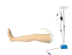 Advanced Venous Infusion Leg Model
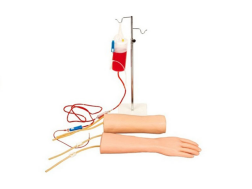 Hand & Elbow Combined IV Training Arm Model


-
Guangzhou, Guangdong, China