
Human Lung Anatomical Model – Pulmonary Bronchial Respiratory System Structure, Medical Teaching Tool for Lung Anatomy and Respiratory System Education

Reference product link:https://www.yddol.com/products/70.html
(Key Features):
Highly Detailed Lung Anatomy
Accurately represents human lung structure, including the bronchial tree, alveolar sacs, lung lobes, and pleura, making it ideal for understanding lung function and pathology.Pulmonary Bronchial System
Features a clear representation of the bronchi, bronchioles, and trachea, showcasing the entire respiratory pathway from the trachea to the alveoli for better comprehension of airflow.Clear Structural Labels
Each part of the model, such as lungs, diaphragm, airways, and blood vessels, is clearly labeled to aid in identification and enhance educational value.Durable & Eco-Friendly Material
Made from high-quality, non-toxic PVC material, the model is durable, long-lasting, and easy to clean, making it suitable for repeated use in classrooms or clinical settings.Removable Components for Interactive Learning
Some models come with removable parts for hands-on teaching, such as lung lobes, bronchi, and vessels, providing students with a better understanding of the organ's construction.
(Applications):
Medical Schools & Universities
Ideal for human anatomy and respiratory physiology lessons, assisting students in visualizing complex respiratory structures.Pulmonary & Respiratory Therapy Training
Perfect for respiratory therapists and pulmonologists to explain lung diseases like asthma, COPD, and pneumonia to patients or students.Emergency Medical Training
Used in first aid, CPR, and advanced life support (ALS) courses to demonstrate respiratory interventions and mechanical ventilation techniques.Patient Education
Excellent for healthcare providers to help patients understand lung diseases, such as chronic obstructive pulmonary disease (COPD), emphysema, or lung cancer.
(Pathologies Represented):
Lung Cancer
Demonstrates the effects of tumors in the lungs, helping in understanding tumor location and treatment approaches.Asthma
Shows the airway constriction and inflammation involved in asthma attacks, aiding in educational explanations.COPD (Chronic Obstructive Pulmonary Disease)
Illustrates how COPD affects the bronchial tubes and alveoli, making it ideal for patient communication about this condition.Pneumonia
Shows inflammation and fluid build-up in the alveolar regions, helping to demonstrate this respiratory infection.Pulmonary Edema
Visualizes fluid accumulation in the lungs, essential for demonstrating heart failure and other related diseases.
(Professional Production):
High Fidelity & Precision: Designed with accurate structural details, ideal for high-level medical training and clinical use.
Interactive & Hands-On: Allows for detailed exploration of the lungs and respiratory system to better understand functionality and pathologies.
The Human Lung Anatomical Model is an essential educational tool for those studying or practicing in the field of respiratory medicine, pulmonology, and general medicine. With its realistic design and clear anatomical representation, this model aids in teaching complex respiratory structures and diseases, ensuring that both students and healthcare professionals can effectively visualize and comprehend the anatomy and function of the human lungs.
 Advanced Arm Blood Pressure Training Model
 Advanced Upper Arm Muscle Injection & Comparison Model w
 Advanced Gluteal Muscle Injection & Anatomical Structure
 Advanced Gluteal Muscle Injection Training & Comparison
 Advanced Electronic Gluteal Injection Training Model
 Advanced Insulin Injection Training Model
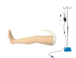 Advanced Venous Infusion Leg Model
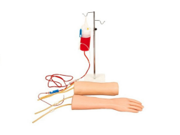 Hand & Elbow Combined IV Training Arm Model


-
Guangzhou, Guangdong, China